
Why Does Puffy Nipple Happen and How To Treat It?
“Puffy nipples” is a term used to describe a certain physical characteristic. It’s where someone’s nipple and areola (the darker skin around the nipple) appear more protruding than...
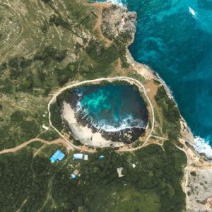
The Ultimate Guide to Angel’s Billabong of Nusa Penida
Nusa Penida, a purely immaculate island at the core of Indonesia, has morphed into a traveler’s paradise looking for solace away from the busy mainlands. Among the many...

The Guide to the Guinea Pig in Peru
Ask any American why you should get a guinea pig, and they will respond that rodents are the cutest pets you can give your children. However, in Peru,...

Everything You Need to Know About Ferragudo, Portugal
Once a remote and lowkey coastal fishing village, Ferragudo is among southern Portugal’s most vibrant tourist towns today. Its natural beauty is incomparable, thanks to its breathtaking orange...
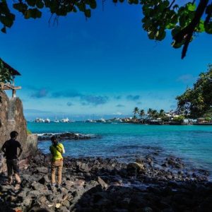
A Complete Visitor Guide to Las Tintoreras, Isabela Island
Las Tintoreras is famous for its scenic treks, mesmerizing marine wildlife, and spectacular snorkeling spots. This small islet within Isabela Island offers a fantastic getaway in one of...

How to Visit the Mermaid Caves on Oahu and How to Get There
Oahu, Hawaii’s third-largest island, is one of the best physical attractions in the world. A fascinating natural phenomenon, the Oahu Mermaid Caves, is among the many riches waiting...
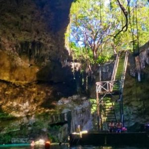
A Complete Guide to Cenote Zaci in Valladolid
In the lovely town of Valladolid in Yucatan, Mexico, I found a wonderful spot named Cenote Zaci Ha. Ever heard of cenotes? They’re sinkholes formed when limestone bedrock...

A Complete Guide to Visiting the Dumbo Manhattan Bridge View
Do you know about New York City’s energy? Well, let me tell you, Manhattan is at its core. A blend of cultures, a hub of arts, and tall,...

109 Fun, Silly, and Meaningful Road Trip Questions for Couples
Need some lighthearted conversation starters to pass the time on a lengthy and exhausting road trip? We’ve got you covered! Here are 109 entertaining road trip questions for...
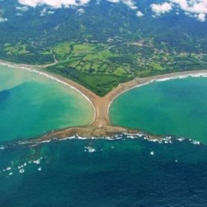
How to Visit the Whale Tail in Costa Rica at Marino Ballena Park
Prepare to go on an adventure of discovery along Costa Rica’s southern Pacific coast. One of nature’s most compelling displays in the region is the Whale Tail. The...